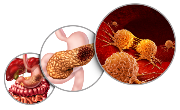
Pancreatic Ductal Adenocarcinoma (PDAC) represents greater than 90% of pancreatic cancer cases [1,2]. Early symptoms are relatively non-specific, with critical early treatment sometimes delayed after misdiagnoses for non-life-threatening conditions such as gallstones [3]. PDAC currently has an average 5-year survival rate of less than 10% [4]. Clearly, there exists a need for better therapy options, with CAR-T immunotherapy being a promising candidate. Potential antigen targets of interest for adoptive immunotherapy against PDAC include: mesothelin (MSLN), CD133, EGFR and HER2.
Mesothelin (MSLN).
Mesothelioma, ovarian cancer, and pancreatic cancer have been found to consistently overexpress MSLN [5]. In pancreatic adenocarcinomas specifically, this overexpression has been confirmed by in situ hybridization, RT-PCR and immunohistochemistry [5]. Non-cancerous pancreatic tissue does not express it or expresses it only weakly. This makes MSLN an ideal antigen for CARs to target. Several Phase I clinical trials have been undertaken involving anti-MSLN CAR T-cells. Limited success has been observed, with the best results being stabilization of disease for between 3.8 and 5.4 months [2]. Ongoing studies are exploring the possible benefits of screening for tumor cell MSLN expression at the outset and using a human-derived rather than murine single chain variable fragment (scFv). Researchers in one study noted the murine origin of their CAR construct may have triggered a host immune response against their modified T cells. Short-lived CAR-T cell persistence was a barrier in all the anti-MSLN trials. This is despite some study participants receiving lymphodepletion preconditioning, in which chemotherapy is administered to kill the patient’s immune cells, prior to CAR-T cell infusion.
CD133.
Aside from PDAC, hepatocellular and gastric carcinomas also express the transmembrane glycoprotein, CD133, though it is present on hemopoietic and epithelial cells more broadly. Nevertheless, due to its high expression in PDAC tumors, at least one anti-CD133 CAR T-cell study explored using the target. Greater than 50% expression of CD133 was detected in the tumor cells of all study participants. Patients were lymphodepleted before receiving two to four infusions of anti-CD133 CAR-T cells intravenously. Positive results included reduction of tumor size, and partial remission or stabilization of disease progression [2]. However, the eventual relapse experienced by study participants may have been due to CD133-negative tumor cells taking over after CD133-positive cells were eliminated. This is termed antigen escape, in which the tumor no longer displays the target antigen for T cells to find. In addition, the microenvironment surrounding tumor tissue presents a number of barriers to infiltration and attack by CAR-T cells. Rather than intravenous administration, as in this study, some studies are exploring locoregional delivery, or infusing the modified T cells via injection directly into the tumor site. This helps get past the extracellular matrix and cancer-associated fibroblasts that often encase tumors.
EGFR.
While this transmembrane protein is associated with tumors, it is also expressed in many other tissues throughout the body. As such, though EGFR is almost always found on PDAC cells, the risk of adverse effects (AE) upon healthy tissue is higher than with more tumor-specific antigen targets. An EGFR-directed CAR T-cell clinical trial was undertaken involving individuals with metastatic PDAC. Though a variety of AEs were experienced by most participants, 75% experienced at least partial response [2].
HER2.
HER2 is a transmembrane glycoprotein that is also present in a number of different tissue types. It is overexpressed on the surface of PDAC cells in 60% of tumors [7]. Use of HER2 as a target faces safety concerns akin to those surrounding EGFR. An anecdotal case-in-point reported is that one study participant passed away within 15 minutes of receiving an anti-HER2 CAR-T infusion. Nevertheless, a subsequent study did see success, relative to that seen in other studies summarized here, though only a minority of participants had a diagnosis of pancreatic cancer. Most participants had biliary tract cancer diagnoses, such as cholangiocarcinoma and gallbladder carcinoma [8]. Despite a variety of AEs seen among study participants, some acute, the two patients with pancreatic cancer diagnoses averaged a period of stable disease of greater than half a year as a result of the anti-HER2 CAR-T therapy [8].
One of the barriers mentioned among these studies is the heterogeneous nature of antigen expression seen in PDAC, as in solid tumors generally. An infusion of CAR-T cells targeting a particular antigen may eradicate all the tumor cells bearing that antigen but those without it will survive and eventually the patient will experience a disease relapse. Looking forward, an area of very promising study is that of multi-targeting CARs. These are T cells modified such that they contain more than one CAR construct and will target multiple antigens, possibly thwarting the antigen-escape route for tumor cells to survive. Studies that have employed multi-targeting CARs have nevertheless still had to contend with low tumor infiltration by CAR-T cells and transient persistence, despite lymphodepleting pre-conditioning. Clearly, numerous barriers presently hinder the advancement of adoptive immunotherapies like CAR T, but the hurdles to be overcome are coming into clearer and clearer view and the promise these therapies hold is a powerful motivator for ongoing research.
References
- Patel, U., Abernathy, J., Savani, B.N., et al. CAR T cell therapy in solid tumors: A review of current clinical trials. eJHaem. 2022;3(Suppl. 1):24–31. doi: 10.1002/jha2.356
- Yeo, D., Giardina, C., Saxena, P., Rasko, J.E.J. The next wave of cellular immunotherapies in pancreatic cancer. Molecular Therapy: Oncolytics Vol. 24 March 2022. doi.org/10.1016/j.omto.2022.01.010
- Ghadimi, B.M, Horstmann, O., Jacobsen, K., Feth, J., Becker, H. Delay of Diagnosis in Pancreatic Cancer Due to Suspected Symptomatic Cholelithiasis. Scandinavian Journal of Gastroenterology, Volume 37, 2002 – Issue 12. doi.org/10.1080/003655202762671323
- Miller KD, Siegel RL, Lin CC, Mariotto AB, Kramer JL, Rowland JH, Stein KD, Alteri R, Jemal A. Cancer treatment and survivorship statistics, 2016. CA Cancer J Clin. 2016;66:271–289.
- Argani, P., Iacobuzio-Donahue, C., Ryu, B., et al. Mesothelin Is Overexpressed in the Vast Majority of Ductal Adenocarcinomas of the Pancreas: Identification of a New Pancreatic Cancer Marker by Serial Analysis of Gene Expression (SAGE). Clin Cancer Res (2001) 7 (12): 3862–3868.
- Yano, Seiichi, et al. “Distribution and function of EGFR in human tissue and the effect of EGFR tyrosine kinase inhibition.” Anticancer research 23.5A (2003): 3639-3650.
- Min, Y., Schwaederle, M., Arguello, D., et al. HER2 expression status in diverse cancers: review of results from 37,992 patients. Cancer and Metastasis Reviews 34 (2015): 157-164.
- Kaichao Feng, Yang Liu, Yelei Guo, Jingdan Qiu, Zhiqiang Wu, Hanren Dai, Qingming Yang, Yao Wang, Weidong Han, Phase I study of chimeric antigen receptor modified T cells in treating HER2-positive advanced biliary tract cancers and pancreatic cancers, Protein & Cell, Volume 9, Issue 10, October 2018, Pages 838–847, https://doi.org/10.1007/s13238-017-0440-4