Biodosimetry & DNA Damage
Visualize and Quantify Inversions with dGH SCREEN™
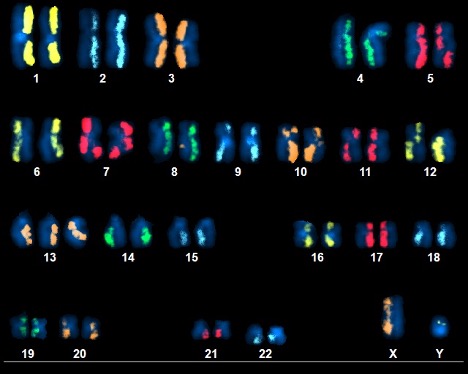
Above Fig.: dGH SCREEN karyogram of metaphase spread from NASA astronaut measuring DNA damage from exposure to the ionizing radiation of space.
The Original KromaTiD Assay.
The original KromaTiD assay, dGH SCREEN was utilized by NASA in the “Twin’s Study” to measure DNA damage from deep space radiation. NASA’s first foray into “omics” research represented a unique opportunity to develop a next-generation approach to measuring the health impacts of spaceflight. Today our dGH SCREEN™ platform is still being used by NASA to understand how to make missions safer for their best and brightest.
dGH SCREEN™ is an unbiased tool for measuring structural and copy-number variants in single-cell fashion. Researchers are able to quantify inversions and other structural variants through direct visualization, an improved biodosimetry method than assaying for the formation of dicentric chromosomes. Inversions occur with lower background frequency than other symmetrical aberrations such as translocations, providing fewer false positives and more reliable data.
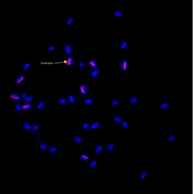
Above Fig.: Murine fibroblasts hybridized with custom dGH biodosimetry paints detecting an inversion event.
Measuring DNA Damage.
DNA damage is a common consequence of exposure to various genotoxic agents, chemicals, and oxidative stress, not just radiation. Understanding the mechanisms of DNA damage and repair is essential for developing strategies to mitigate the adverse effects of genotoxic insults. Many model systems serve to further such research, such as rodents.
This image features the chromosomes of a mouse fibroblast used as part of a biodosimetry investigation. dGH tools can be applied in non-human settings as well. Based on synthetic oligonucleotides, custom dGH probes can be manufactured to target sequences in other published mammalian genomes. This includes rodents, larger mammals like dogs and cats, or commercial lines like CHO cells. As demonstrated in this image, the dGH system can successfully detect structural variants outside of the human genome setting.
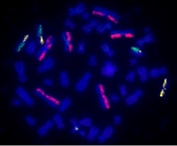
Above Fig.: Edited cell hybridized with dGH SCREEN whole-chromosome paints showing translocations, signal gain, and an inversion or sister-chromatid exchange.
Quality Control for Gene Editing.
Gene editing can be inherently destabilizing to the genome, especially when multiple edits are carried out. However, for many applications like CAR-T cell engineering, the therapeutic cell product requires more than one genomic locus to be modified. While dGH SCREEN can be applied in a whole-genome fashion, the individual tool components are dynamic. A subset of the dGH SCREEN paints may be used, depending on the dataset desired. This may be done in a targeted way, painting only those chromosomes impacted directly by the editing. Alternatively, a set of larger chromosomes may be painted as a proxy with which to gather data on the overall rate of structural rearrangements and off-target effects. In this way, the data generated can be tailored to the needs of the editing project.