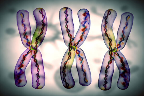
Optical Genome Mapping (OGM) is a technology which employs microfluidics, image analysis, and high-resolution microscopy to detect structural variations (SVs). The process begins with the isolation of high molecular weight DNA from biological samples. This DNA is then fluorescently labeled, counterstained, and loaded onto a chip. This is not a sequencing chip. Rather, individual DNA strands are run through pores to gather structural data from the pattern of fluorescent labels along the length of each strand. While a powerful tool for detection of chromosomal aberrations, recent research1 has revealed some of the limitations of OGM. Investigators report circumstances under which the technology is prone to false negatives, and recommend having a thorough understanding of its limitations, especially when it is applied in clinical settings.
As can be the case with sequencing platforms, the errors predominantly arise where the breakpoints of chromosomal aberrations are located in regions rich in repetitive DNA sequences, such as segmental duplications (SDs). SDs are long, repeated sequences estimated to represent more than six percent of the human genome.2 Repetitive regions pose a challenge for technologies which involve a sequence alignment step. The algorithms employed can miscalculate the location a DNA segment when more than one location has the same sequence. OGM does not work by aligning DNA sequences but faces this dilemma as well because it aligns the pattern of fluorescent labels present on each DNA strand.
Researchers report the choice of reference genome does influence the quality of OGM results. Through a data accuracy comparison using GRCh37 or GRCh38 as reference genomes, data revealed varying detection rates for chromosomal aberrations. While use of GRCh38 assembly produced fewer false outcomes, both produced an error rate near five percent. Increasing reading depth was not found to improve these results.
Despite its limitations, OGM remains a valuable tool, especially when used in conjunction with other technologies to generate orthogonal datasets. Single-cell platforms without an alignment step, such as directional Genomic Hybridization (dGH™) SCREEN can help fill in the gaps left by OGM. dGH SCREEN analysis examines the full chromosome complement, one cell at a time through direct visualization. This means errors in the alignment step can be corrected through visual confirmation. G-band karyotyping provides some of the same benefits but can only resolve features larger than about 10 megabases. dGH SCREEN can achieve a limit of detection as low as 10 kilobases.
It is clear that a broad assessment of any genomic tool is warranted before use, weighing considerations such as false-data vulnerability, cost, throughput, sample-type limitations, and the level of expertise required to process resulting data. No genomic analysis platform is infallible, and the most reliable scientific conclusions are reached through the joining of complementary techniques. Understanding the conditions under which each tool may generate questionable results is essential for informed decision-making in clinical and research settings.
Reference:
1. Xu, Y., Zhang, Q., Wang, Y., et al. (2024). Optical Genome Mapping for Chromosomal Aberrations Detection—False-Negative Results and Contributing Factors. Diagnostics, 14(2), 165. doi.org/10.3390/diagnostics14020165
2. Pu, L., Lin, Y., & Pevzner, P. A. (2018). Detection and analysis of ancient segmental duplications in mammalian genomes. Genome Research, 28, 901-909. doi:10.1101/gr.228718.117