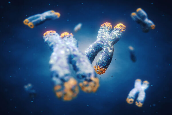
DNA damage is occurring within our cells constantly. Like a boat that is springing new leaks all the time, repair must also be ongoing or the ship will sink. Organisms have thus evolved check and repair mechanisms to undo this continuous injury. However, this creates its own problems. The ends of our chromosomes risk looking a lot like the end of a broken DNA strand. Signaling mechanisms must be in place for the cell to decide what to do about them. If it decided to “repair” the seemingly broken DNA, it might join it with another chromosome end with potentially deadly consequences for the cell. The cell must know to do nothing. This is the purpose of the telomeres, which are special repeating sequences on the ends of our chromosomes that say to that cellular machinery that this DNA isn’t damaged. Leave it alone. These DNA regions are the subject of great interest because they shorten as we age, losing just a little bit of length with each cell division. Eventually, this shortening threatens the ability of telomeres to communicate that “stand down” message to those proteins involved in the cell’s DNA repair tasks. That in turn, risks telomere-initiated senescence, a permanent stop to the cell cycle. The alternative is apoptosis, the death of the cell. Here, we discuss one method that exists for measuring the length of a cell’s telomeres: Quantitative Fluorescence In Situ Hybridization (Q-FISH).
Fluorescence In Situ Hybridization (FISH) is a well-established method by which nucleic acid molecules bearing fluorescent tags result in a spectral signal wherever they locate and bind another nucleic acid molecule whose base pair sequence is complementary. Standard FISH is generally considered semi-quantitative, which is where Quantitative-FISH, Q-FISH diverges.
Q-FISH telomeric probes are complementary to the repetitive DNA of the telomeres, a series of repeating segments of the sequence, TTAGGG. These probes must be as short as possible but still functional. They represent the measuring rod which will be used as the base unit of telomere length, so shorter probes result in a higher-resolution measurement. Individual probe molecules will produce a cumulative change in signal brightness as their total grows. However, the quantitative nature of the method comes about thanks to the controls put in place. Fluorescent beads with known brightness are included in the reaction mixture. These, along with the imaging hardware and software, enable scientists to correct against variation in the illumination intensity and other sources of noise. By this system, units of telomere length can be counted via an incremental change in fluorescence signal intensity.
Telomere length studies are at the root of investigations into ageing, cancer and even the health risks inherent to space travel. Discoveries made within this arena have the potential to lead to products and therapies for extending the normal human lifespan. Q-FISH is just one of many methods employed in furtherance of this goal.